Patients with painful spinal stenosis – compression of the nerves in the back or neck – can gain relief from their suffering without enduring the physical insult of open surgery and the attendant healing time.
An effective treatment comes through a tiny incision the size of a button. And the procedure sometimes can be done on an outpatient basis, so patients go home the same day.
As with any other, it takes practice for a surgeon to become adept at this technique. The University of Louisville School of Medicine has at its disposal a facility that now is making this practice easier for neurosurgeons in training. At its fresh tissue lab, residents can practice on cadavers before they take their skills to the operating room.
The lab has been a resource for other departments well since 1981, but the neurosurgery department had never made use of the lab. That changed on Friday, Oct. 16.
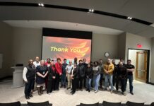
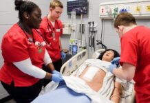
Under the direction of new neurosurgery department chair Jonathan Hodes, M.D., the five UofL neurosurgery residents gowned up and first watched, then tried, the minimally invasive procedure during a three-hour workshop.
We’ll make a smaller incision and use telescoping dilaters to spread, rather than cutting and burning tissue, Hodes said as he began to demonstrate the technique. Later, each of the residents got to try it.
There’s not another place in the vicinity that has this kind of capability, said Andy Dzenitis, M.D., a UofL professor of neurosurgery and auditor of the workshop.
In addition to serving as a training resource for current residents, the lab has been an attractive recruiting tool for new trainees in other disciplines, including otolaryngology, obstetrics and gynecology and orthopedics, said Robert Acland, M.D., lab director.
The fresh tissue dissection laboratory is among the few nationally recognized facilities dedicated solely to the highest caliber training of surgeons, said Susan Tate, M.D., fellowship director for female pelvic medicine and reconstructive pelvic surgery in the Department of Obstetrics, Gynecology and Women’s Health. Our female pelvic reconstructive surgery program attracts superior fellows in large part due to our association with this unique facility.
The lab, said Hodes, is a precious resource for all lifelong medical learners because it allows, in a compassionate, respectful and safe environment, for learners at all levels to hone and develop surgical skills with no risk to patients. This laboratory exercise is an essential part of residency training and helps the school of medicine to create more skilled and better prepared neurosurgeons for our community.
One of the largest and best-equipped facilities for fresh tissue dissection in the United States, the facility provides unique educational opportunities for learning and re-learning anatomical relationships as well as pioneering new surgical techniques in a safe environment, said Fred Roisen, Ph.D., chair of the department of anatomical sciences and neurobiology, which administers the lab.
In addition to training residents, the undergraduate and graduate academic departments at UofL have used the lab for national and international professional education programs.
For their part, the neurosurgery residents who used the lab said the experience was beneficial.
The cadaver lab was a good learning experience, said Abdul Baker, M.D., a third-year neurosurgery resident. We got hands-on exposure to new spine equipment and to minimally invasive spine technology.


